Diagnostic imaging plays a crucial role in modern oncology, providing physicians with the necessary tools to effectively detect, diagnose and treat cancer. These advanced technologies not only improve diagnostic accuracy, but also enable detailed monitoring of treatment progress, contributing significantly to improved patient outcomes.
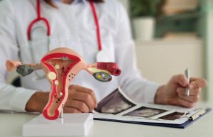
Diagnostic imaging uses a variety of technologies to create detailed images of the inside of the body, allowing physicians to observe and analyze organs and tissues without the need for surgery. These images are essential for detecting the presence of tumors, determining the extent of disease and planning appropriate treatment.
Types of diagnostic imaging in oncology
- Computed Tomography (CT): Uses X-rays and computer technology to create detailed 3D images of the body. CT is especially useful for detecting tumors, assessing the extent of cancer and guiding biopsy procedures.
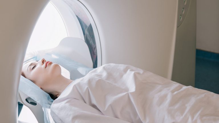
- Magnetic Resonance Imaging (MRI): Uses magnets and radio waves to produce detailed images of soft tissues. MRI is ideal for imaging the brain, spinal cord and other areas where accuracy is critical.
- Positron Emission Tomography (PET): Combines nuclear medicine and CT to show metabolic and biochemical activity in the body. PET is effective in detecting cancer, assessing its extent and monitoring response to treatment.
- Ultrasound: Uses sound waves to create real-time images of internal organs. Ultrasound is commonly used to guide biopsies and evaluate tumors in the abdomen and pelvis.
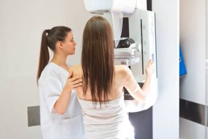
- Mammography: A specific X-ray technique used to examine breast tissue and detect breast cancer in its early stages.
- X-ray: Uses x-rays to visualize the bones and detect bone lesions or bone metastases.
Benefits of diagnostic imaging in oncology
- Early detection: Imaging techniques make it possible to detect cancer in its early stages, when it is more treatable and the chances of cure are greater.
- Diagnostic accuracy: Detailed images provide critical information about the location, size and extent of the tumor, facilitating accurate diagnosis and treatment planning.
- Treatment monitoring: Imaging allows physicians to evaluate the effectiveness of ongoing treatment, adjusting plans as necessary to maximize results.
- Surgical planning: Detailed images guide surgeons during procedures, ensuring that as much cancerous tissue as possible is removed while preserving healthy structures.
- Follow-up and relapse detection: Diagnostic imaging is essential for the long-term follow-up of patients, detecting any cancer recurrence early.
El diagnóstico por imágenes es una herramienta indispensable en la oncología moderna, proporcionando la precisión y el detalle necesarios para detectar, diagnosticar y tratar el cáncer de manera efectiva. Si tienes preguntas o necesitas más información, no dudes en contactarnos.
Specialists:


Dr. Humberto Juarez
Cardiovascular Surgery / Thoracic Surgery / Endovascular Surgery / Minimally Invasive Surgery
See Specialist